
Congenital Conditions
Foot Pathologies
CLUBFOOT
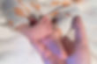
Clubfoot (or congenital talipes equinovarus – CTEV) is a common birth defect where one or both of a baby’s feet are twisted inward. Around 50% of cases affect both feet.
There are three types: postural, idiopathic, and syndromic. Postural clubfoot is usually mild and may resolve without treatment. The idiopathic type (most common) and syndromic clubfoot generally require medical care.
Most cases of idiopathic clubfoot are treated with the Ponseti method, which uses a series of weekly plaster casts to gently correct the foot. In some cases, a minor tendon release (tenotomy) is needed.
Once corrected, the baby wears a clubfoot brace (foot abduction brace) to maintain alignment and prevent relapse. When started early and followed properly, this treatment is highly effective.
CONGENITAL VERTICAL TALUS (CVT)
Congenital Vertical Talus (CVT) is a rare but serious foot deformity in newborns where the foot bones are misaligned. Instead of the navicular bone sitting properly on the talus, it shifts upward, forcing the talus into a vertical position.
This creates a visible rocker-bottom foot deformity — the sole curves outward, the heel points down, and the front of the foot lifts up. Unlike flexible flat feet, CVT is stiff and rigid, and does not correct on its own.
Parents may notice a convex foot shape or a rigid flatfoot shortly after birth. CVT can occur alone or be linked to neuromuscular conditions like spina bifida or arthrogryposis. It’s important to know that CVT is not caused by anything parents did during pregnancy.
Early treatment is key. It typically starts with gentle stretching and serial casting (like the reverse Ponseti method) to gradually align the foot. In many cases, a minor surgery is needed, followed by bracing to maintain correction.
With timely and proper care, most children with congenital vertical talus grow up to walk and play normally.
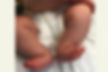
TALIPES CALCANEOVALGUS

Talipes calcaneovalgus is a common and usually harmless foot condition in newborns, where the baby’s foot appears bent upward and outward, with the heel pointing downward. This newborn foot deformity often results from the baby's position in the womb — known as a packaging defect — especially when there’s limited space.
At birth, the baby's foot may look deformed, but the condition is typically flexible and not painful. In most cases, it improves on its own with gentle stretching exercises done at home. Calcaneovalgus foot in babies usually resolves within a few weeks to months without surgery.
However, if the foot remains stiff or does not improve, physiotherapy may be needed. In rare cases, persistent deformities are evaluated further to rule out underlying neuromuscular conditions.
HIP PATHOLOGIES
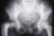
DEVELOPMENTAL DYSPLASIA OF THE HIP (DDH)

Developmental Dysplasia of the Hip (DDH), also known as Congenital Dislocation of Hip, is a condition where a baby’s hip joint is unstable or improperly formed, ranging from mild looseness to complete dislocation. Key risk factors for hip dysplasia in babies include family history, breech position during pregnancy, being female, and first-born babies. Parents may notice signs like a hip click in baby, uneven leg length, or difficulty with crawling or walking. If untreated, DDH can lead to hip pain, early arthritis, limping, knee and back pain, and an abnormal walking pattern. Early diagnosis, often via hip ultrasound for newborns or hip screening, is essential. When detected early, many cases respond well to non-surgical treatment with a Pavlik harness or hip brace for DDH, avoiding surgery. In cases diagnosed later, surgery for developmental dysplasia of the hip may be needed to promote normal hip development and prevent long-term complications. Parents often ask, does DDH go away on its own? Early intervention offers the best chance for a healthy, pain-free life.
DEVELOPMENTAL COXA VARA
Developmental Coxa Vara is a rare hip deformity in children where the angle between the head and shaft of the femur (thigh bone) is abnormally decreased, causing the top of the femur to tilt inward. This condition often leads to a shorter leg, a noticeable limp, and sometimes hip pain or limited movement in young kids. It usually becomes clear when a baby or toddler starts walking. The exact cause of coxa vara is often unknown but may relate to abnormal growth of the upper femur. Parents may notice an abnormal gait in their child or a leg length difference due to coxa vara. Diagnosis is confirmed through a child’s hip X-ray. While mild cases may only require observation, many need surgical correction for coxa vara to restore proper hip alignment, improve leg length, and prevent long-term joint problems. Physical therapy can also help in some cases. Parents often ask if coxa vara will get better on its own, but early diagnosis and treatment are important for the best outcome.
KNEE PATHOLOGIES
CONGENITAL DISLOCATION OF THE KNEE (CDK)

Congenital Dislocation of the Knee (CDK) is a rare newborn knee deformity where the knee joint is abnormally positioned at birth, with the lower leg (tibia) bent backward in relation to the thigh (femur). Parents may notice that their baby's knee is bent backward or that the knees don't bend properly. This condition often results from tight or shortened quadriceps muscles and may be linked to other conditions like arthrogryposis or occur alongside clubfoot and knee dislocation.
Signs of knee dislocation in newborns are usually visible at birth and confirmed with a baby knee X-ray. CDK can range from mild to severe. Early treatment is essential and often starts with gentle stretching, physiotherapy, and casting for congenital knee dislocation. In more severe or resistant cases, surgery may be required to release tight structures and properly align the joint.
Parents often ask, "Will my baby walk normally with knee dislocation?" — and the good news is that, with timely intervention, most children achieve good knee function and mobility over time.
LIMB DEFICIENCIES
CONGENITAL FEMORAL DEFICIENCY (CFD)

Congenital Femoral Deficiency (CFD) is a rare birth condition where the thigh bone (femur) is either shorter or underdeveloped, leading to a leg length difference, hip or knee instability, and challenges with walking in children. It is not caused by anything parents did during pregnancy—it occurs during early development in the womb. Parents may notice one leg is shorter than the other, a visible limp, or a baby born with a short thigh bone. Sometimes the foot may appear smaller or turned outward, and fibular hemimelia may also be present.
Diagnosis of femoral deficiency in babies usually happens in infancy or early childhood through physical exams and X-rays. Treatment depends on severity and can involve limb lengthening for CFD, beginning with hip and knee stabilization surgeries such as the SUPER Hip and SUPER Knee procedures. Gradual leg lengthening is done using external fixators or intramedullary nails, and in some cases, growth modulation of the longer leg may be used to help achieve balance.
Each femur deficiency treatment plan is personalized based on the child’s anatomy, function, and long-term mobility goals. With expert care, many children with congenital femoral deficiency go on to walk and thrive.
FIBULAR HEMIMELIA
Fibular Hemimelia is a rare congenital limb deformity where a child is born with part or all of the fibula bone missing—the smaller bone in the lower leg. Parents may notice a baby born without fibula, a short leg, or a foot that looks small or curved. This condition often leads to a leg length difference, ankle and foot deformities (like a turned-in foot or missing toes), and knee instability. Fibular deficiency can range from mild underdevelopment to complete absence of the fibula and is usually diagnosed at birth or sometimes through prenatal ultrasound.
Though the exact causes of fibular hemimelia are unknown, it is not related to anything done during pregnancy and typically isn't inherited. Treatment for fibular hemimelia is highly personalized and may involve SUPER ankle reconstruction, knee stabilization procedures, and limb lengthening using external fixators or other advanced techniques. In some cases, guided growth may be used to manage growing limb length differences. For more severe cases, parents may explore amputation vs. lengthening as part of long-term planning.
The goal is to ensure your child can walk, run, and live confidently, with or without a prosthetic leg, depending on the best option for their anatomy and function.
RADIAL CLUB HAND

Radial Club Hand, also known as Radial Longitudinal Deficiency, is a rare congenital arm deformity where the radius bone in the forearm is underdeveloped or missing. This causes the baby’s wrist to curve inward, bending toward the thumb side (radial side) of the forearm. In some cases, the baby may be born with a bent wrist, a short or curved arm, or even a missing thumb. The condition can range from mild shortening to a completely absent radius and may affect one or both arms. Radial deficiency in newborns is sometimes associated with syndromes such as VACTERL.
Signs of radial club hand are often visible at birth and include a curved forearm, wrist deformity in newborns, or a baby's hand turned inward. Treatment for radial club hand depends on the severity and usually begins early with stretching, splinting for curved wrist, and in more severe cases, surgical correction to improve wrist alignment and hand function. With the right orthopedic treatment for radial deficiency, many children gain good hand use, improved appearance, and the ability to carry out daily activities independently.
Dr. Gourav Jandial is a highly skilled Paediatric Orthopaedic Surgeon specializing in non-surgical clubfoot treatment and complex foot deformities like Congenital Vertical Talus (CVT). Trained at multiple international institutes including University of British Columbia, the Paley Orthopedic & Spine Institute, University of San Fransisco and others with experts such as Dr. Kishore Mulpuri, Dr Anthony Cooepr, Dr. Dror Paley and Matthew Dobbs, he offers advanced, evidence-based care for conditions like Club foot (CTEV), Developmental Dysplasia of the Hip (DDH) and Radial Club Hand. Experienced in limb reconstruction and managing rare deformities including Fibular Hemimelia and Congenital Femoral Deficiency, Dr. Jandial develops personalized LIFE PLANs that guide treatment from infancy through growth, aiming to preserve natural limb function and improve quality of life. His global training and research contributions, along with compassionate family support, ensure children receive the best care for active, independent lives.
His academic contributions include presentations at national and international conferences, such as:
"Controversies in Tibialis Anterior Transfer in Relapsed Clubfeet: Split/ Total; Cuboid/ Cuneiform" – JPOA Conference 2019, Osaka, Japan
"Complications of Tibialis Anterior Transfer in Clubfoot: A Single Centre Study" – Global Clubfoot Conference 2017, New Delhi, India
"Correlating the Bilateral Feet in Idiopathic Clubfeet" – IOACON 2017, Indore, India
Single-Event Versus Staged Open Reduction in Bilateral Developmental Dysplasia of the Hip: Does Rate of Early Dislocation Vary? Canadian Orthopaedic Association Annual Meeting, Halifax, Nova Scotia, June 2024.
Tethering In Sleeper Plate Mode For Guided Growth: Does The Plate Material Matter? - EPOS Meeting 2025, Toulouse, France
A Novel Technique To Prevent Stump Overgrowth In Skeletally Immature Patients With Traumatic Crush Injury Of Foot/ Leg - POSICON 2024, Guwahati, India
Limb Lengthening And Reconstruction Society (LLRS) Aim Index – Reliability In Assessing Disease Severity - LLRS Meeting 2023, Lake Tahoe, USA